Brain Cancer Scientists
Spencer Gibson, PhD
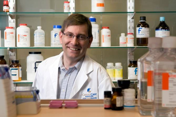
In maintaining integrity and homeostasis of multicellular organisms, the balance between cell death and survival is fundamentally important. When this balance is altered, diseases such as cancer occur. One protein important in the regulation of cell death is BNIP3 which is induced under low oxygen (hypoxia) conditions and is over expressed in solid tumours. This paradox of BNIP3 killing cancer cells while being over expressed in live cells within tumours is a focus of our research. Three explanations could account for these differences and act as a mechanism for cancer progression.
Cell survival is as important as cell death. The epidermal growth factor receptor (EGFR) is expressed at high levels in several cancers including breast cancer. We discovered that pretreatment of breast cancer cell lines with epidermal growth factor (EGF) effectively blocked drug and death receptor induced apoptosis. This protection from apoptosis is mediated by a serine threonine kinase (AKT) through up-regulation of the Bcl-2 anti-apoptotic family member Mcl-1. Besides breast cancer, we have found that a lipid, lysophosphatic acid (LPA) blocks apoptosis in chronic lymphocytic leukemia (CLL) cells using a similar mechanism. We are currently investigating the regulatory elements controlling Mcl-1 expression.
The goal of my research is to define the signal transduction pathways leading to cell death or survival. This will elucidate targets that could tip balance in favour of cell death and will be the foundation to establish clinical trials using molecular targeted therapies to increase effectiveness of chemotherapy in cancer.
Contact Information:
5008b-675 McDermot Ave
Winnipeg, Mb
R3E 0V9
Phone: 204-787-2051
Email: Dr Spencer Gibson PhD
Dr Sachin Katyal, PhD
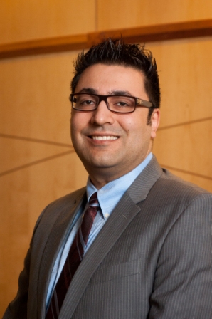
Modulating DNA Damage repair mechanisms to enhance brain tumour treatment success
DNA strand breaks occur on a daily basis in cells due to cell stress, environmental factors, oxidation and metabolism. Damaged DNA is resolved by dedicated DNA damage response (DDR) and repair mechanisms in order to preserve genomic integrity and cell function. The goal of conventional chemotherapeutic drugs and radiotherapy is to elicit DNA damage to overwhelm the tumour’s innate DDR and induce cell death. However, tumour cells have remarkable ability to respond to DNA damage, repair and adapt thus allowing survival and eventual drug resistance. It is predicted that >90% of all tumours incur at least one defect in the DNA damage response (DDR), thus tumour cell survival relies upon enhanced activity of other compensatory DNA repair pathways. The aggressive and deadly brain tumour, glioblastoma multiforme (GBM) shows a very high level of recurrence due to emergence of chemo/radio-resistant tumour cell populations; patients usually live about 1 yr from their date of diagnosis. We are identifying the “back-up” DNA repair pathways in these deadly brain tumours so that we can enhance the patients’ treatment success and their quality-of-life.
DNA repair pathways are guardians of the cellular genome.
Every individual human cell is estimated to incur tens of thousands of DNA strand breaks due to environmental stress, oxidation, metabolic function and DNA decay. To preserve genomic integrity, these breaks are resolved by dedicated DNA damage repair (DDR) pathways that ensure faithful transmission of genomes in dividing cells to ensure proper cell function and survival. There are two classes of DNA strand breaks, double-strand breaks (DSBs) and DNA single-strand breaks (SSBs), which are resolved by specific repair pathways, DSBR and SSBR. The inability to properly process and repair SSBs can interfere with the DNA replication and transcriptional machinery resulting in persistent SSBs, formation of the particularly genotoxic DNA double-stranded break (DSB) lesion and aberrant gene expression resulting in a variety of cellular pathology, including: senescence, cancer and apoptosis. It is known that the diverse mechanisms involving cell cycle regulation, DDR pathways, cellular metabolism, and cell death act in concert in response to DNA damage. As such, cellular life and death decisions are balanced by these mechanisms as defective DDR in proliferating cells, including neuroprogenitors, can lead to cancer, while defective neuronal DDR can lead to neurodegeneration.
Anti-tumorigenic agents overwhelm cellular DNA repair responses with lethal levels of genotoxicity.
The objective of common front-line radiation and chemotherapeutic strategies used in the treatment of brain tumours is to induce DNA breaks so as to overwhelm the cellular DNA repair machinery thus promoting genomic damage and tumour cell death. However, as the intrinsic cellular DNA repair process counteracts the therapeutic efficacy of this strategy, high radiation and drug doses are required which result in harmful neural and systemic side effects. My research seeks to identify ways to dysregulate cellular DNA repair pathways in tumours and improve therapeutic success. In this regard, DNA damage repair pathways are an ideal clinical target as we can specifically kill cancer cells by lowering the radio- and chemotherapeutic threshold of tumour cell genotoxicity by inhibiting redundant DNA repair pathways.
My research uses advanced molecular, biochemical and genetic techniques to gain insight into the biology of mammalian DNA strand break repair pathways. My goal is to identify ways to manipulate these pathways to develop novel treatment strategies in the clinical management of cancer. We are seeking a very highly motivated postdoctoral fellows (with a strong history of previous success) to join our team.
Contact information:
675 McDermot Ave
Winnipeg, Mb R3E 0V9
ph.: 204-787-2765, fax: 204-787-2190
Email: Dr Sachin Katyal PhD
Dr. Thomas Klonisch, MD PhD
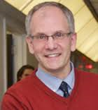
I am interested in understanding mechanisms tumor cells utilize to bypass anticancer treatment in patients and use this information to develop more effective anticancer therapies. This includes testing the efficacy of novel small molecules, evaluating synthetic lethal combinations, and the use of nanotherapeutic strategies. Although I utilize mice for this research, such animal models have limitations for some human cancers, in particular brain cancer. We are employing methods to isolate brain tumor cells directly from patients and have generated an extensive brain tumor cell resource. This invaluable resource allows my translational team to test therapeutic strategies and address important questions related to gliomagenesis, stem/ progenitor cell plasticity, cellular heterogeneity within human brain tumors, survival strategies, and mechanisms of tissue invasion and metastasis of adult brain tumors. In addition, we employ brain tumor cells and patient blood samples to identify potential protein biomarker signatures and monitor immunological responses in glioblastoma patients.
Two molecular systems of great interest to me are the membrane-anchored G protein coupled relaxin receptor system and the nuclear stem cell factor High Mobility Group A2 (HMGA2) in brain and breast cancer cells. My team established a role for the relaxin receptor RXFP1 in human glioblastoma and identified a novel ligand of RXFP1 in human brain tumors. Using our established resource of isolated human glioma cells from human patients, my team was able to show a novel mechanism by which this RXFP1 receptor system mediates chemoresistance to temozolomide in patient brain tumor cells. My research on the chromatin-binding protein HMGA2 has revealed important roles of this stem cell factor in human patient glioblastoma cells. We showed that HMGA2 is a base excision repair (BER) enzyme and protects replication forks and telomeres. We also demonstrated that HMGA2 alters the activation state of the ATR-CHK1 DNA repair signaling which protects GB cells from apoptosis upon exposure to DNA alkylating agents like temozolomide. Currently, we study the interaction of HMGA2 with key proteins involved in cell survival and investigate proteins and pathways we identified as synthetic lethal with HMGA2. Collectively, our results suggest that the RXFP1 receptor system and HMGA2 exert novel protective mechanisms at different cellular levels which collectively result in enhanced chemoresistance in cancer cells. Our ongoing research addresses two main goals: (i) identify protein signatures from patient blood samples that can indicate the presence of glioblastoma and (ii) use sophisticated basic science strategies to inform the development of more efficacious treatment options for cancer patients.
Information:
Dr. Thomas Klonisch
Professor & Head
Dept. of Human Anatomy and Cell Science
Director of the Histomorphology and Ultrastructural Imaging Platform
Director of the Glioma Cell Resource
Depts. of Surgery, Medical Microbiology & Infectious Diseases
Adjunct Scientist, CancerCare Manitoba
Honorary Professor of Shantou University Medical College, Shantou, China
Max Rady College of Medicine, Rady Faculty of Health Sciences, University of Manitoba
133-745 Bannatyne Avenue, Winnipeg, Manitoba, Canada, R3E 0J9
Phone: +1 204 789 3893
Fax: +1 204 789 3920
Email: thomas.klonisch@umanitoba.ca
Dr. Tamra Werbowetski-Ogilvie, PhD

Central nervous system (CNS) tumors are among the most prevalent forms of childhood cancers accounting for nearly 20% of all new cases (Canadian Cancer Society Statistics, 2015). Medulloblastoma (MB) is the most common malignant primary pediatric brain tumor and is currently divided into 4 distinct molecular subtypes.: WNT, SHH, Group 3 and Group 4. Extensive genetic, molecular and clinical heterogeneity between these subgroups has made it difficult to assess the functional relevance of genes to tumor progession. The Werbowetski-Ogilvie lab employs high throughput flow cytometry-based screening platforms to identify and subsequently characterize novel roles for cell surface markers in regulating diverse medulloblastoma phenotypes both in vitro and in vivo. Utilizing a wide variety of functional assays including measures of self-renewal, differentiation, invasion and proliferation in vitro as well as tumor growth in vivo using xenograft models, the lab is currently investigating the role of CD271, also known as p75 neurotrophin receptor (p75NTR) or nerve growth factor receptor (NGFR), in regulating stem cell properties of SHH variant medulloblastoma cells.
In addition, the lab utilizes neural derivatives from human embryonic stem cells (hESCs) as a model system to study the mechanisms contributing to pediatric brain tumorigenesis. Using this powerful cell resource as both a complement to and surrogate for existing cell lines and heterogeneous patient samples, the goal is to better understand how genes such as the transcription factor orthodenticle homeobox 2 (OTX2) regulate the balance between self-renewed and differentiation to either prevent or sustain oncogenic properties.
Contact Information:
University of Manitoba
611-745 Bannatyne Avenue, BMSB
Winnipeg, MB R3E 0J9
Phone: 204.789.3431
Fax: 204.789.3900
Lab: 204.977.5687
Tamra.Ogilvie@umanitoba.ca
Dr. Marco Essig
Dr. Marshall Pitz
Dr. Marshall Pitz is the Section Head of Clinical Research at the Research Institute at CancerCare Manitoba (CCMB). He conducts clinical research on brain, breast, and lung cancers, as well as health services research on the use of information technology in the delivery of cancer care. Dr. Pitz is also a medical oncologist at CCMB where he treats patients with brain and breast cancers; an Associate Professor of Medicine in the University of Manitoba’s Rady Faculty of Health Sciences; and CCMB’s Chief Medical Information Officer responsible for the clinical use of the electronic medical record.
Research
Dr. Pitz leads a research team testing an advanced type of magnetic resonance imaging (MRI) to improve the treatment of patients with glioblastoma, the most aggressive type of brain cancer. Conventional MRIs cannot reliably distinguish brain tumour progression from a phenomenon called pseudo-progression, which may be brain tissue inflammation that is a positive response to treatment. If the advanced MRI technique is more effective at distinguishing true tumour progression from pseudo-progression, it will allow Dr. Pitz and other oncologists to identify patients who require additional treatments, and spare other patients from aggressive (and potentially harmful) surgeries and/or chemotherapy they are unlikely to benefit from.
Dr. Pitz is a member of a multi-disciplinary research team evaluating physical therapy and acupuncture as treatments for a disabling side effect of chemotherapy (peripheral neuropathy) in breast cancer patients.
Using tissue specimens from the Manitoba Tumour Bank, Dr. Pitz led a research team that examined how androgens (steroid hormones) may affect the treatment outcomes of patients with non-small cell lung cancer, the most common type of lung cancer.
Publications
Dr. Pitz has published over 25 peer-reviewed papers since 2002, including one of the most frequently cited 2014-2018 papers with a Research Institute scientist as its lead author.
Dr. Pitz’s publication list (PubMed)
Contact Information
Phone: (204) 787-2197
E-mail: mpitz@cancercare.mb.ca
Dr. Lawrence Ryner
Dr Magimairajan Issai Vanan, MD, MPH, FAAP

Our lab’s main focus is in translational Neuro-Oncology with special emphasis on Radiation and Chemotherapy sensitization of pediatric brain tumors, developmental neurobiology as related to brain tumors and neuro therapeutics (BBB permeability / novel drug delivery methods).
Radiation is an integral part of the therapeutic armamentarium in Pediatric Neuro-Oncology. The therapeutic benefits of radiotherapy are, however, accompanied by late toxicity that severely affects quality of life in children. Our lab has identified several potential targets that mediate radiation and chemotherapy resistance in pediatric brain tumors. Validation of these targets using patient derived xenografts (PDX) in orthotopic murine brain tumor models will provide us with novel radio-sensitization drugs with larger therapeutic window; when used with current treatment protocols, this may lead to low dose therapeutic radiation and less long term side effects in survivors of childhood brain tumors.
The blood vessels that vascularize the central nervous system (CNS) possess unique properties, termed the blood–brain barrier (BBB), which allow these vessels to tightly regulate the movement of ions, molecules, and cells between the blood and the brain. However the same barrier also prevents chemotherapeutic drugs from reaching the tumor. The BBB is frequently intact in diffuse intrinsic pontine glioma (DIPG) and restricts the delivery of systemically administered conventional and biological therapies. In collaboration with Dr Miller we are working on chemical (HAV / Cyclic peptides) and biological (SHH and Wnt) methods of transient alteration of BBB permeability in orthotopic tumor models (DIPG). This transient increase in BBB permeability will help us increase the efficacy of current therapy and hopefully improve outcome in this devastating tumor.
Contact information
Dr Babu Sajesh PhD.
ON5025a-675 McDermot Ave
Winnipeg, MB, R3E 0V9, Canada.
Ph: 204-787-4171
For more information please contact:
CancerCare Manitoba
Phone: (204) 787-2197
Toll-free: 1-866-561-1026
Copyright © 2000-2019 CancerCare Manitoba.
All Rights Reserved.
 Accreditation Canada")